About this episode
You can listen and download our episodes for free on more than 10 different platforms:
https://linktr.ee/dentistry_made_simple
Produced by:
https://www.podcaistudio.com/
Get every episode summarized
Each time Dentistry Made Simple publishes, we email you a written briefing from the transcript — the topics, who appeared, and any specific claims, with the ad reads skipped.
Email me new episodesFree for 3 shows. No card needed.
Hosts & guests
Transcript ready
285 searchable segments. Every word is indexed and playable.
Full transcript
Dentistry Made Simple — Endodontic Radiology. Machine-transcribed; use the interactive transcript above to jump the player to any line.
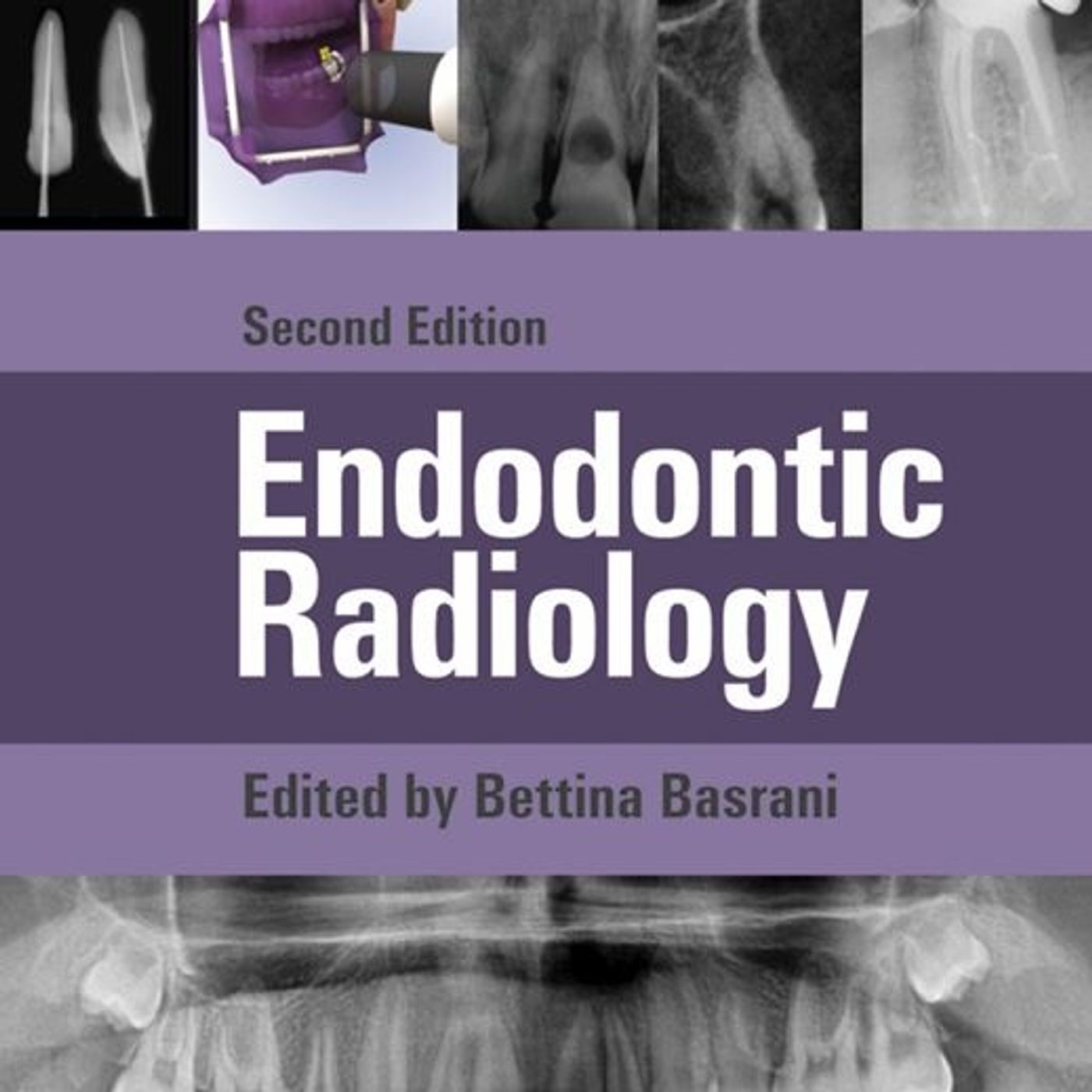
you are operating inside the microscopic caverns of a toothroot entirely surrounded by dense bone. Yeah, completely blind. Exactly. You are essentially flying blind. Yet somehow as a clinician, you are expected to navigate this microscopic terrain with absolute pinpoint precision. I mean, there's a sheer magic to the field of endodontics when you stop and really think about it. Oh, absolutely. It is the ability to peer into these hidden dark zones of human anatomy and heal what we absolutely cannot see with the naked eye. Well, it is a profound paradox for any clinician, right? You are tasked with curing a disease process that you cannot physically look at. Right. So imaging isn't just like a helpful supplementary tool. It is the entire foundation of the discipline. I mean, without it, endodontic therapy is just guesswork in the dark. Just poking around. Exactly. Because if you cannot see the root architecture, you cannot clean it and you certainly cannot heal it. Okay, let's unpack this. We are basing today's deep dive on a really foundational pillar of the field. It's the textbook endodontic radiology second edition,
which is edited by Dr. Bettina Bizarani. A fantastic resource. It really is. And our mission today is to give you our listener, whether you're a dental student or a young professional, a complete, highly detailed roadmap of endodontic imaging. Yeah, we're covering a lot of ground today. We are. We are going to start in the incredibly dangerous early days of X-rays, dig into the physical limitations of the 2D techniques you use every single day and then explore the massive 3D revolution of cone beam computed tomography. Sounds great. But, you know, to really appreciate the pristine digital sensors and modern safety protocols we have today, we first have to understand the genesis of the glow. Genesis of the glow. I like that. We really need to acknowledge the literal physical sacrifices that were made to bring X-rays into dentistry. The timeline, it really begins back in 1870. Okay, 1870. Right. There was a physicist named Wilhelm Hitorff. And he found that if he passed an electrical discharge through a partially evacuated glass tube. Like a vacuum tube. Yeah,
exactly. He noticed it emitted these mysterious rays that caused a greenish yellow glow. So people were noticing these weird glowing effects in the lab, but they didn't really know what they were looking at yet. Precisely. The monumental shift happened decades later on November 8, 1895. Right. Ronbian. Yes. Wilhelm Roenchen. He was experimenting in a darkened room and realized the same type of tube was emitting an invisible ray that actually caused crystals all the way across the room to fluoresce. Just spontaneously lighting up. Yeah. He called them X-rays with the X standing for the unknown. And to prove his discovery, he exposed his wife's hand to these rays to capture the shadows of her bones. Okay, I have to ask because we're so used to modern X-rays taking what? A fraction of a second. Yeah, millisecond. Right. So how long did she actually have to sit there while this highly experimental machine just bombarded her hand? 15 solid minutes. Oh wow. Yeah, a massive dose of raw radiation by today's standards. And the dental pioneers, they were right on his
heels. Just 14 days after Rankin's discovery, a German dentist named Otto Wachoff made the very first dental radiograph. Just two weeks later. Just two weeks. He wrapped a glass photographic plate in black paper and rubber, put it in his own mouth, and submitted himself to 25 minutes of continuous X-ray exposure. 25 minutes of raw, un-sultured radiation straight to the face. Yeah. I mean, it sounds like the Wild West of science. It really was. Just an era of incredible boundary pushing discovery, but fatal recklessness. Fatal is the right word. What's fascinating here is how incredibly dangerous it was simply because there was a complete lack of biological understanding. They just didn't know. Right. And that danger is perfectly encapsulated by a dentist from New Orleans named Dr. C. Edmund Kells. In 1896, Kells became the first practitioner to use intrural X-rays on a live patient. And by 1899, he was using them to actually determine the length of a root canal during therapy. But here's the tragic part. Kells exposed his hands to X-rays every single day for years. Why is hands? Well,
he would physically hold the glass plates inside his patient's mouths to adjust the bean quality while the machine was running. Wait, he was just using his own bare hands to stabilize the film while the machine was actively firing. Yes. Every single day. And that unprotected exposure eventually led to aggressive cancer in his hand. Oh, no. Yeah, it resulted in the amputation of his fingers and then his hand and eventually his entire arm. That is horrific. It was a brutal, tragic demonstration of the biological price of ionizing radiation. Was anyone sounding the alarm back then or is everyone just blasting themselves with radiation in the name of progress? There was one distinct voice of reason, actually. In that exact same year, 1896, a Boston dentist named William Rollins published the first paper on the severe dangers of X-rays. For him? Yeah, he was way ahead of his time. He proposed filtering the beam, collimating it. Collimating meaning, like, restricting the beam. Exactly. Restricting the size and shape of the X-ray beam so it doesn't just scatter everywhere. And he even suggested covering the patient with a lead apron. In 1896, that's basically our modern
protocol. Exactly. But it took years for the actual technology to catch up to his ideas. It wasn't until 1913 that William Coolidge developed a high vacuum tungsten tube that finally stabilized the machine's output. Wow. Let's talk about the biological mechanism behind all this because it's vital for a clinician to really understand. Biologically speaking, why exactly does radiation target certain tissues over others? That's a great question. Like, why did kels get cancer specifically in his skin while other tissues in his hand might have survived that kind of daily exposure? The precise mechanism comes down to cellular division. You see, ionizing radiation fundamentally damages DNA. It can actually sever the strands of the DNA double helix. This snaps them. Right. And this damage is most catastrophic during mytotic division. That's when the cell is unzipping its DNA and actively dividing. Okay, so when it's most vulnerable? Exactly. Radiation increases the incidence of DNA defects during this vulnerable phase. And it actively interferes with the body's normal ability to repair those defects. Because
of this, tissues whose cells divide frequently are vastly more sensitive to radiation damage. Okay, so we are talking about rapidly turning over cells being the primary targets here. Yes, exactly. Highly susceptible cells include hematopoietic cells. So the ones in your bone marrow that are constantly churning out new blood cells. Right. It also includes immature reproductive cells, young bone cells, and epithelial cells like the skin. Ah, the skin. Right. On the flip side, cells that divide very slowly or not at all are considered radiation resistance. So mature bones, muscles, and nerves fall into this category. That makes total sense. So the epithelial cells on KELS hands were constantly divided to regenerate his skin, making them incredibly vulnerable to the DNA shattering effects of his daily exposures. Exactly. It's a chilling context, really. It is. It really makes you realize why today our use of radiation is governed by such strict agencies relying heavily on the ICRP, the International Commission on Radiological Protection. Yes,
and they hammer home the AALARIO principle. Right. Keeping radiation doses as low as reasonably achievable. AALARIO. So assuming a clinician is following a LARA, the next step is actually applying this safely. Standard 2D radiography is basically the absolute foundation of the diagnostic process. It has some pretty severe physical limitations that you absolutely must understand to avoid misdiagnosis, right? Yes. Let us first establish what a clinically acceptable correct radiograph should actually achieve. Okay, let's go over the criteria. You use radiographs to view the root canal space, spot periapical lesions at the tip of the root, identify fractures, and determine your working length. Standard bread and butter stuff. Exactly. But to be considered a high quality diagnostic image, it must meet very strict criteria. First, it has to capture the full root plus at least two millimeters of the periapical bones surrounding the apex. Right, you need that border of bone. Second, if there is a pathology like a lesion, the complete rare faction must be visible. Rare refaction, meaning the entire dark area of bone loss. Correct.
The whole dark area plus a border of normal healthy bone around it. Third, it needs minimal distortion, and fourth, it needs optimal density and contrast. And if you miss those marks, you end up with defective radiographs, which are, frankly, entirely non-diagnostic. Exactly. They're useless. The literature outlines four major errors you'll encounter. Can we explain how these errors actually happen on a physical level? Because I know a standard film relies on silver-helid crystals. That is the perfect way to understand it, actually. Traditional radiographic film is coated in a gelatin emulsion containing silver-halid crystals. Okay. When x-ray photons strike these crystals, they sensitize them. Then later, during chemical development, those sensitized crystals are converted into black metallic silver. So more x-rays mean more black silver? Exactly. The more x-rays that hit an area, the darker that area becomes. This explains our first error, the under-exposed image. Right. The one that looks too light. Right. This happens because the exposure time was too short,
or maybe the development time was insufficient, meaning not enough crystals were converted to silver. Got it. Then you have the overexposed image, which is way too dark. And that is the exact opposite. Too much radiation time, or too much time in the chemical developer converting way too many crystals. Spot on. Then there's the blurred image, which is easily recognized by fuzzy borders. That's just someone moving, right? Yeah. A simple mechanical failure. The patient, the film, or the x-ray tube moved while the beam was active. Finally, we have the partial image, frequently called a cone cut, or a collimation error. Ah, cone cut. Remember we mentioned earlier that collimation restricts the beam's shape? Yeah, the keep it focused. Well, if you don't properly align that restricted central beam directly over the film, a section of the film simply doesn't get hit by x-rays at all. Oh, leaving that curved, unexposed blank spot on your final image. I love looking at the physics of it. But, you know, here's the major problem. Even if you take a mathematically perfect, beautifully processed 2D
radiograph, you are still fighting against physical reality. You are. Looking at a 2D radiographs, kind of like looking at a shadow puppet of a dog on the wall. You can clearly see the outline of the ears and the snout, but you have no idea how the hands and fingers are actually twisted together to make that shape. That is a brilliant analogy. Thanks. I mean, you are completely blind to the depth of the object. Yes. The 2D shadow completely misses the buckle-lingual dimension. Buckelingual. Right. Which refers to the depth from the cheek, the buckle side to the tongue, the lingual side. You are taking a complex three-dimensional anatomical structure and flattening it into a single flat plane. Which has to create massive diagnostic blind spots. Huge blind spots. For instance, a radiolucin lesion, a dark spot indicating bone loss, will not even appear on a periapical radiograph unless a substantial amount of bone has already been destroyed. Wait, let me make sure I understand this. Significant, canceless bone resorption. So the destruction of the spongy, honeycomb-like bone deep inside the jaw that won't even register
on a 2D x-ray. It will not. That's wild, why not? Because the dense outer layer of the jaw bone, the cortical bone, is so thick that it basically masks the loss of the spongy bone inside. The x-ray beam still hits enough dense calcium in that outer layer to show up white on the film. You will only see the dark spot of a lesion once the infection has eaten all the way through the spongy bone and actually resorbed that dense outer cortical plate. So the absence of a dark spot on your film does not mean there isn't an active boneding infection happening right in front of you. Exactly. It doesn't eat far enough yet. And even when you do see a dark spot, the 2D film cannot reliably tell you what kind of tissue replace the bone, right? Not at all. I mean a 2D shadow cannot differentiate a fluid-filled ridiculous cyst from a solid dental granuloma or even freeing healing scar tissue. And this raises an important question about how we interpret these images. Because this limitation directly causes a major issue with observer bias. Radiographic interpretation is
notoriously subjective. Everybody sees something different. Exactly. There is a classic study by Goldman that beautifully illustrates this. Researchers had different endodontists and radiologists look at the exact same set of recall radiographs to evaluate the success or failure of various root canals. Let me guess they couldn't agree. The results were alarming. The experts disagreed on the diagnosis more often than they agreed. Wow. Yeah. The 2D image simply leaves way too much room for human interpretation. Right. Because the 2D shadow hides that crucial cheek to tongue depth. And because you need massive bone loss just to see a lesion, the field absolutely had to evolve. We needed a better view. We needed a way to see the hands making the shadow puppet, basically. And this brings us to the massive 3D paradigm shift. Cone beam computed tomography or CBCT. Yes, CBCT. It was introduced to dentistry in 1998 and it completely revolutionized diagnostics. How does it differ from a regular medical CT scan? Well, traditional medical CT
take multiple flat slices and expose the patient to very high doses of radiation. CBCT is different. It uses a cone-shaped x-ray beam that makes a single 180 to 360 degree rotation around the patient's head. Just one quick spin. Right. In that one sweep, it captures a cylindrical or spherical volume of data called the field of view. And the detail it produces is staggering, right? I've seen the specs on these machines. We are talking about voxelsizes ranging from 0.08 to 0.4 cubic millimeters. Yes, and it's really important to understand what a voxel actually is. Okay, break it down for us. Think of a standard pixel on your TV screen. It is a flat 2D square. A voxel is a volumetric pixel. It is a 3D cube. When you build an image out of microscopic cubes, as small as 0.08 millimeters, you create a flawless digital replica of the tooth. This allows the clinician to slice through the tooth in all three anatomical planes simultaneously. Let's give everyone a mental map of those planes because it's so crucial for navigation in 3D. Of course,
first, the axial plane. That is a horizontal slice, like looking down at the tooth from above, almost like a drone shot. Got it. Then the coronal plane. That's a vertical slice from side to side, like looking at the tooth face on in a mirror. And finally, the sagittal plane is a vertical slice from front to back, looking at the tooth from its side profile. And because you can view all these angles, CBCT just completely eliminates superimposition. Where one anatomical structure, blocks another. That's exactly. Which means you can detect pariapical disease far earlier than conventional 2D films. You don't have to wait for the dense outer cortical bone to be destroyed. You can actually see the spongy bone breaking down inside. It is also revolutionary for evaluating highly complex root canal anatomy. Like what? I imagine a C-shaped canal, which is a treacherous ribbon-like web of anatomy. On a 2D film, it often just looks like a standard root. But on a CBCT, you can see every intricate curve. It is also essential for planning apical microsurgery, where you need to know exactly how many
millimeters lie between the root tip you're cutting and the mandibular nerve. It sounds like an absolute magic bullet. But what's the catch? I know there's a specific map reading strategy clinicians use, but the hardware itself has limitations, right? Artifacts are a major headache. Oh yes. Artifacts can severely hinder a CBCT scan. If a patient has an intracanol metallic post or heavily radiopech crown materials, it actually scatters the cone beam. The bounce is it everywhere. Right. The software gets confused by the dense metal and creates these massive white streaks known as starburst artifacts. Oh starbursts. Yeah. And these streaks can completely obscure the root structure you are trying to examine, rendering the 3D scan basically useless for that specific tooth. But when it works without artifacts, the diagnostic power is undeniable. There's a brilliant clinical example in the text regarding anatomical mimics that I want to talk about. Oh yes. The nasopalatine ductsist. Right. Imagine you take a 2D radiograph of a maxillary central incisor cell,
one atop front teeth. You see a well circumscribed dark circle right at the tip of the root. It looks exactly like apical periodontitis, like a classic root infection. And if that tooth already had a root canal, your first instinct is that the treatment failed, and you need to open the tooth back up. Exactly. But with the CBCT scan, you can view that exact same area from the side in the sagittal plane. And you will clearly see that the dark circle isn't attached to the tooth apex at all. It's just sitting behind it. Right. It is actually a nasopalatine ductsist sitting far behind the tooth deeper in the palate. On the 2D film, the shadow of the cyst was simply superimposed over the tooth apex, which is incredible. The CBCT completely changes the diagnosis and saves the patient from a highly invasive, completely unnecessary endodontic retreatment. If we connect this to the bigger picture, it perfectly proves that the shadow puppet can lie to you. Yeah. This level of clarity is actually forcing the profession to rewrite it's standards of success. For decades, researchers used the pariapical index or P.A.I., which was developed by Brian Alff and
and that was based on 2D films, right? Completely based on 2D films. It was a scoring system for apical period contitis. But now, CBCT is forcing researchers to completely re-evaluate those traditional success and failure rates. Peace we're seeing more. We're suddenly seeing failing lesions that were always there. They were just hiding in the buckle, lingo blind spot, the 2D shadow. Okay, here is where I really have to push back, though. Okay, go ahead. If CBCT is this vastly superior, incredibly detailed 3D map, why are we still using 2D? Like, shouldn't CBCT replace 2D entirely for every single patient that walks through the door? Why even bother with the shadow puppet anymore? It is a really tempting thought, but the answer is a resounding no. Really? No. No. Conventional 2D imaging, specifically using the paralleling technique to minimize distortion, that remains the mandatory starting point for routine cases. We must always return to the alair app principle. As low as reasonably achievable. Exactly. Even though CBCT uses far less radiation than a medical CT, it still exposes the patient
to more radiation than a standard digital pariapical radiograph. Oh, okay. That makes sense. You also have to consider the high cost of the equipment availability, and frankly, the sure amount of time it takes to painstakingly analyze hundreds of slices in a 3D volume. You only deploy CBCT when the initial 2D image cannot answer your diagnostic question. Got it. It's a specialized, targeted tool, not a blunt instrument. So CBCT is the current clinical pinnacle for live patients. But how do researchers study the actual histology of these lesions without extracting and destroying teeth? And for that matter, how do you train a dental student to understand 3D anatomy before they ever touch a live patient? Well, let's explore alternative imaging first. Ultrasound uses high resolution transducers to capture real-time echoes based on the acoustic properties of the tissues. Like sonar. Kind of, yeah. It sends high-frequency soundways into the jaw. When those sound waves hit a boundary between different materials like solid tissue versus fluid, they bounce back as an echo. The machine translates those echoes into an image. Oh wow. And the massive advantage here
is that it uses zero ionizing radiation. Zero. And because it's mapping acoustic density, it can do something CBCT struggles with, right? Exactly. Because it maps acoustic echoes, ultrasound can uniquely differentiate between a solid granuloma and a fluid-filled cyst. How does it do that? By detecting vacuillization. That's the actual fluid-filled spaces within the lesion. That's brilliant. It is. But the caveat is that sound waves transfer heat and mechanical energy into biological tissues. This is a process called cavitation. So a heat's up the bone. Basically. Therefore, the exam time must be strictly limited to prevent thermal damage to the surrounding cells. So it's a balance. But we have micro CT, which sounds like CBCT's hybrid-detailed cousin, used strictly for research, right? Yes. Micro CT is used exclusively for in-veature research on extracted teeth. It is based on the original CT principles developed by Sir Gottfried Hounsfield. Electrical engineer. Right. The guy who co-invented the CT standard. Micro CT provides phenomenally accurate, non-destructive 3D images. In endodontic research,
this is how we actually evaluate our tools. Okay, walk me through that. Well, a researcher stands in extracted tooth, uses an endodontic file to clean the canal, and then stands it again. Before and after. Exactly. The software overlays the two 3D models. The negative space perfectly highlights exactly which microscopic parts of the canal wall were left entirely untouched by the instruments. That is exactly how we proved that round files don't perfectly clean oval-shaped canals. Precisely. But what about the students? I mean, you can't put a multi-million dollar micro CT in a pre-clinical lab. Yeah. How do you teach a student to see that hidden third dimension when all they have is a basic 2D x-ray machine and a plastic typeodont model? Oh, there is an incredibly clever pre-clinical exercise designed specifically for this. Okay, love this one. The instructor takes a conventional piece of dental film and folds it in half, placing a thin piece of lead foil right in the middle crease. The student places an extracted tooth on one half of the folded film and takes an x-ray. Then they flip the folded film over to
the unexposed side. They change the angle of the x-ray tube, shifting it either measly, meaning toward the front midline, or distally, poured the back, and take a second shot of the exact same tooth. And because of the lead foil in the middle, the radiation from the first shot doesn't bleed through and ruin the second half of the film. Precisely. When they develop that single piece of film and unfold it, they have two different views of the exact same tooth sitting side by side, taken from two different horizontal angles. That is so cool. It forces the student's brain to mentally integrate those two 2D shadows to build a complex internal 3D model of the root canal system. It is essentially an analog hack to teach a digital era 3D concept. It really is. It highlights just how utterly essential spatial awareness is for a clinician. You have to build that 3D model in your mind's eye. And that is the ultimate takeaway here. From Roachin and Kells exposing themselves to raw radiation without understanding the biology, to the frustrating, overlapping shadows
and physical limits of 2D x-rays, right up to the crystal clear diagnostic power of CBCT, ultrasound, and microCT. The evolution is just breathtaking. It is. But you know, the technology doesn't make the diagnosis you do. Radiology is still, in many ways, more of an art than a strict science. Without a doubt. An image, no matter how high resolution the voxels are, is only a tool. True, accurate diagnosis always requires integrating these advanced radiographic findings with physical clinical tests. I mean, a massive dark shadow on a CBCT means absolutely nothing without a simple cold test to confirm if the pulp tissue inside the tooth is actually dead or alive. Exactly. So to reinforce everything we've covered today, here is a short clinical exercise for you, the listener, to visualize. All right, laid on us. Imagine you are looking at a 2D periapical radiograph of a maxillary central incisor. This tooth has had previous root canal treatment. You spot a distinct, well circumscribed radioleucency right at the apex. Okay, got the picture.
But when you talk to the patient, they are completely asymptomatic. No pain, no swelling. Based on what we discussed today regarding anatomical mimics and the limits of the 2D shadow, what specific nonendodonic pathology must you include in your differential diagnosis before you even consider opening that tooth for retreatment? You have to consider the nasal palatine ductsist. It is the classic anatomical mimic in the anterior maxilla, hiding right there in the buckle angle blind spot. Spot on. Keep that diagnostic skepticism in your mind as you step up to the chair tomorrow morning. But I want to leave you with one final thought to mull over. Okay. Something looking past the current era of CBCT. If human observer bias is still our biggest hurdle, if experts still disagree on what a shadow means, how long until we aren't the ones doing the interpreting? That's a wild thought. Right. Imagine an artificial intelligence trained on millions of micro CT and CBCT scans. An AI that doesn't suffer from eye fatigue that can instantly detect a shift of a single 0.08 millimeter voxel, flaking a microscopic failure before the human eye
can even perceive it. The x-ray might pierce the darkness, but very soon it might not be a human mind that actually sees the light.
More episodes
More from Dentistry Made Simple

Endodontics: Principles and Practice
Dentistry Made Simple

Emergency Medicine Oral Board Review: Pearls of Wisdom
Dentistry Made Simple

Facial Aesthetics: Concepts and Clinical Diagnosis
Dentistry Made Simple

Essentials of Pharmacology for Dentistry
Dentistry Made Simple